
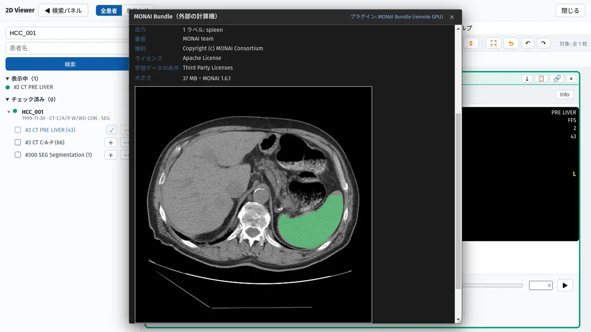
MONAI Bundle
Runs MONAI Bundle segmentation models on an external GPU such as Google Colab and loads the labels into the ROI manager and DICOM SEG.
Get it on GitHub ↗A desktop edition that works entirely offline, and a web edition that connects to an external PACS. One interface covering 2D, 3D and quantitative analysis.
For research use only — not for diagnostic use
Stack display, window/level presets, cine, and intensity calibration (HU and SUV managed together).
Reslicing in three orthogonal planes, with gantry tilt correction, plus MIP, MinIP and AvgIP slab projection in 2D, MPR and 3D.
Oblique planes at any angle and three kinds of curved planar reformation along a centreline.
Volume and surface rendering, cinematic rendering, 3D measurement and clipping.
Draw and manage 2D regions of interest, paint masks, and convert between ROIs and meshes.
Texture analysis through RadiomicsJ, with a settings interface.
Fusion overlays and SUV calibration (body weight and others).
Storage, DIMSE, DICOMweb and REST, query/retrieve, and remote send.
A plugin mechanism, with a Japanese and English interface.
3D and MPR are rendered on the GPU with WebGL2, including cinematic rendering (Monte Carlo path tracing). If the GPU cannot be used and rendering falls back to software, GRAPHY-Next detects it and tells you.
GRAPHY-Next keeps its core small and adds capability through plugins, so you install only what each analysis needs.
Public Exclusive Public: free, available on GitHub. Exclusive: provided only to site-support (enterprise plan) customers. Apply for site support →
Runs MONAI Bundle segmentation models on an external GPU such as Google Colab and loads the labels into the ROI manager and DICOM SEG.
Get it on GitHub ↗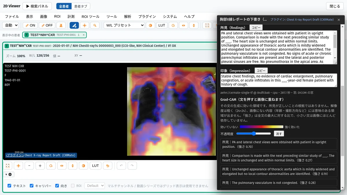
Drafts findings and an impression for one chest radiograph with CXRMate, on this PC only. The text can be wrong and is not for diagnosis.
Get it on GitHub ↗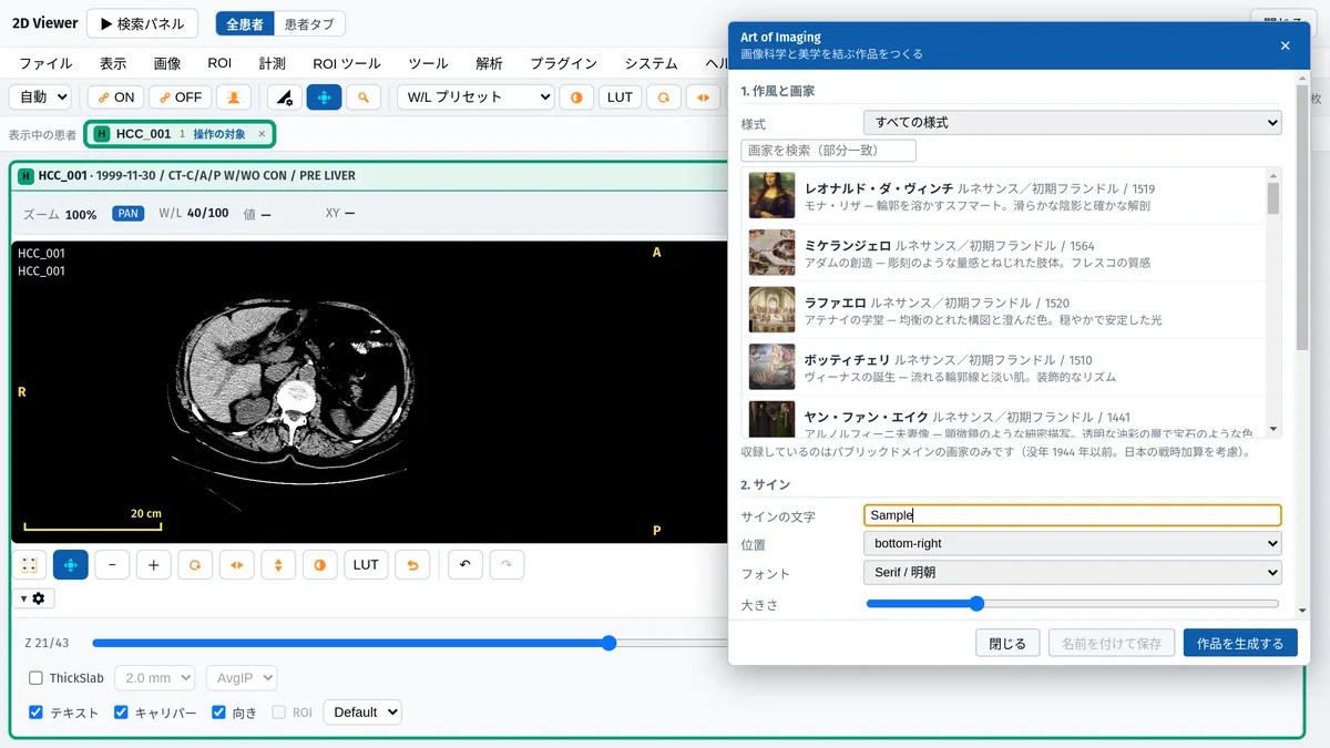
Turns the displayed image into an artwork in the style of a public-domain painter, with an appreciation note. For artistic expression, not diagnosis.
Get it on GitHub ↗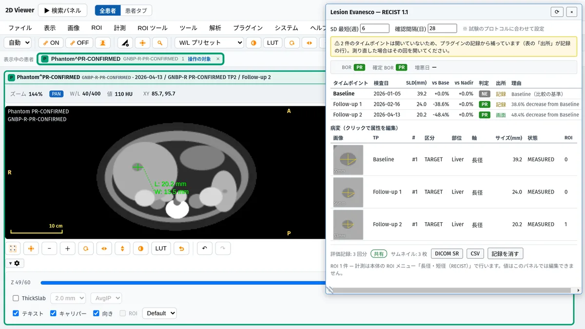
From diameters measured with the built-in ROI tools, assesses response per time point, best overall response (BOR) and progression date.
Apply for site support →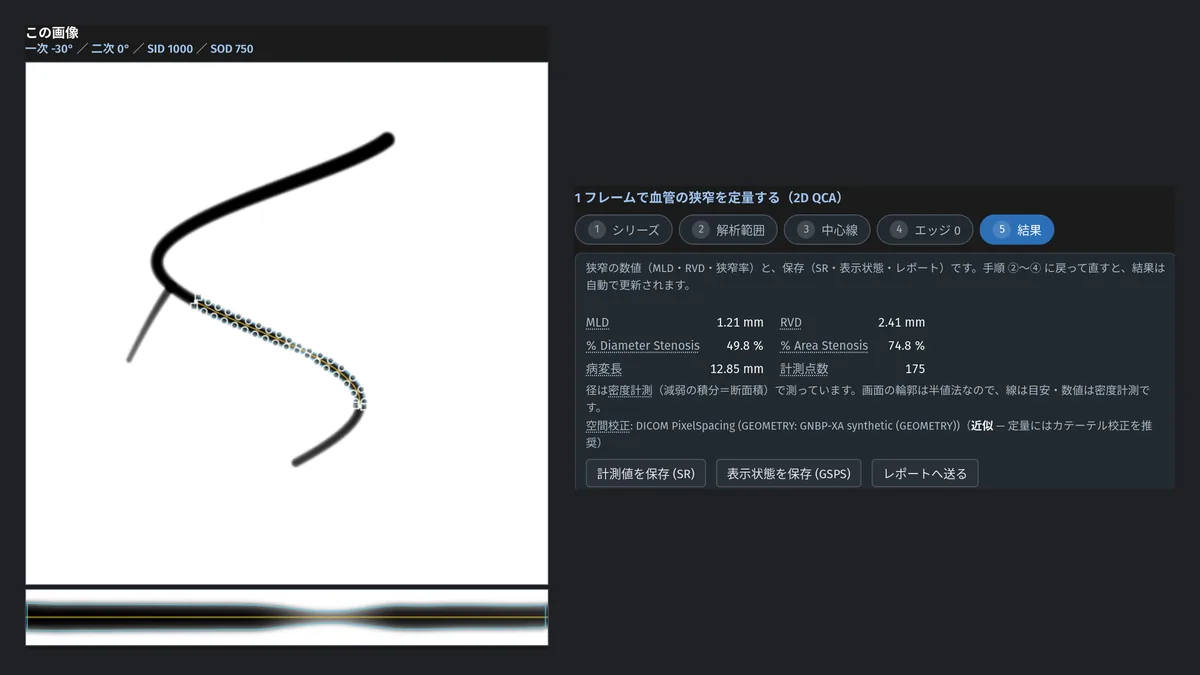
Quantitative angiography from XA runs: 2D QCA, QLV, 3D QCA (single vessel and bifurcation) and QFR.
Apply for site support →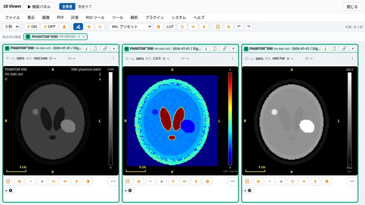
From DWI at several b-values, generates ADC, computed DWI (cDWI) and a diffusion-derived virtual MRE.
Apply for site support →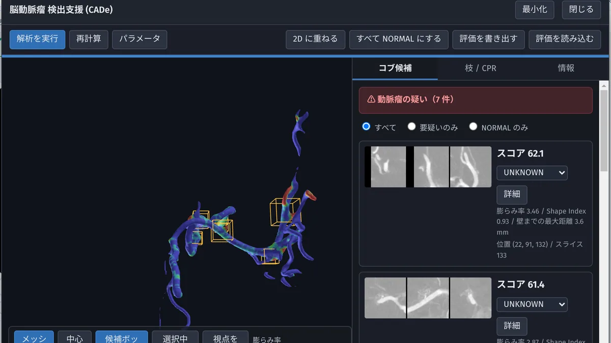
Extracts the cerebral vessel centerline and branches and flags aneurysm candidates. A reading aid, not a diagnosis.
Apply for site support →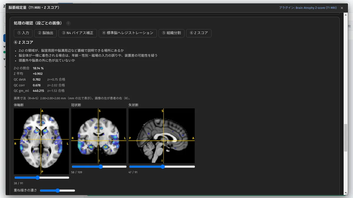
Builds a gray-matter atrophy Z-score map against a healthy-reference database from a 3D T1 MRI, with a report, on this PC only.
Apply for site support →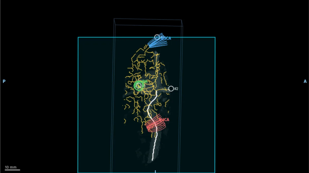
Quantifies blood flow in the basilar and both internal carotid arteries from 4D Flow MRI and compares it with 2D PC.
Apply for site support →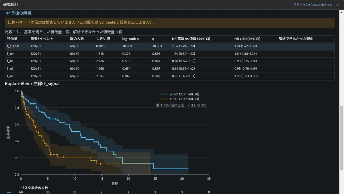
Joins radiomics features with clinical data into a dataset and runs published analysis skills.
Apply for site support →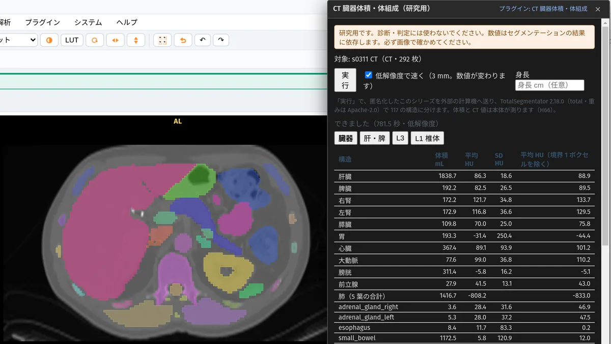
Segments organs with TotalSegmentator on an external GPU; the app measures volume, CT values and L3 muscle area.
Apply for site support →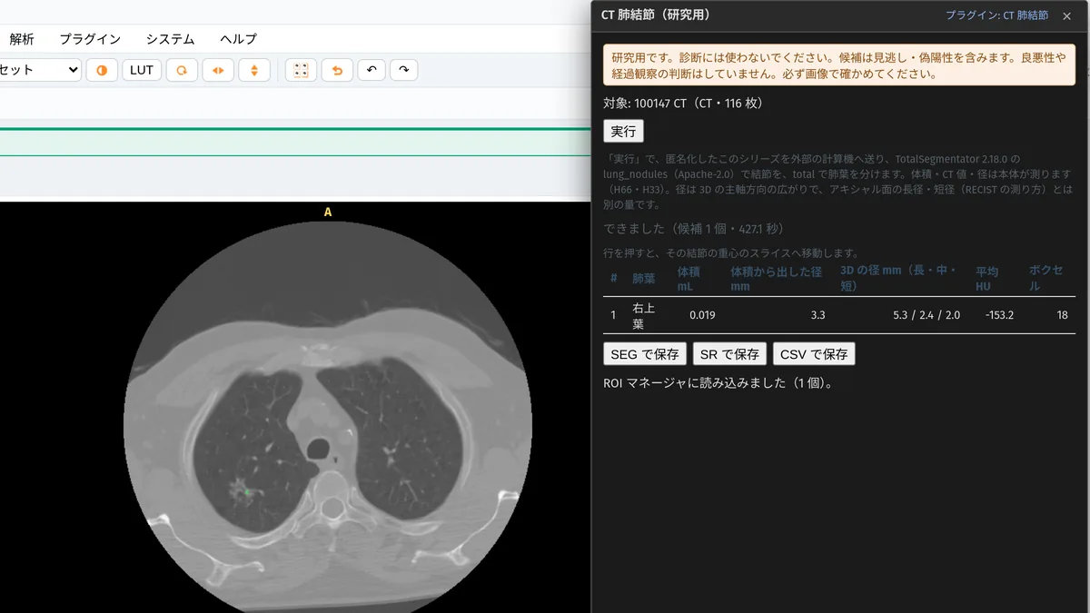
Lists nodule candidates with TotalSegmentator on an external GPU, with lobe, volume and diameter. It does not classify malignancy, and can miss or over-call.
Apply for site support →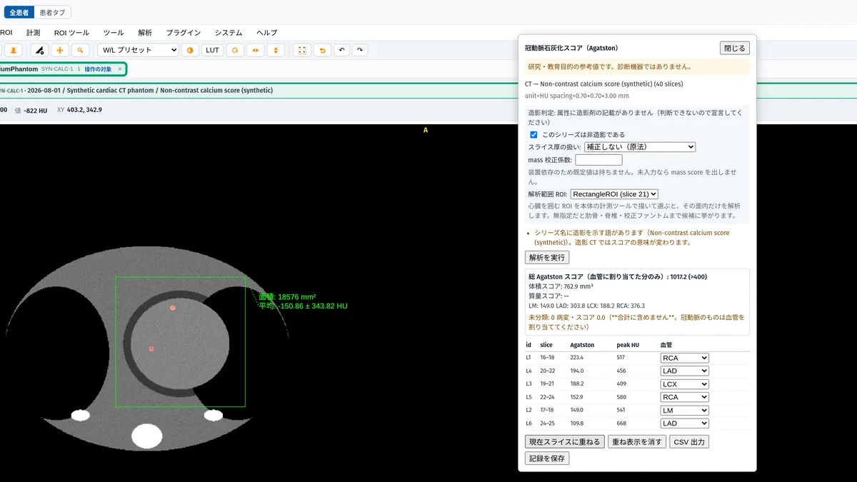
Quantifies calcium score, late gadolinium enhancement (LGE), T1/T2 and ECV, and the AHA 17-segment model.
Apply for site support →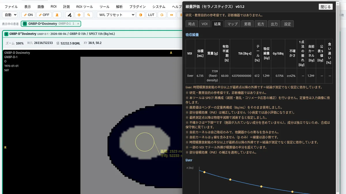
Patient-specific absorbed dose for Lu-177 therapy, from time-activity curves to dose-volume histograms. Not for treatment planning.
Apply for site support →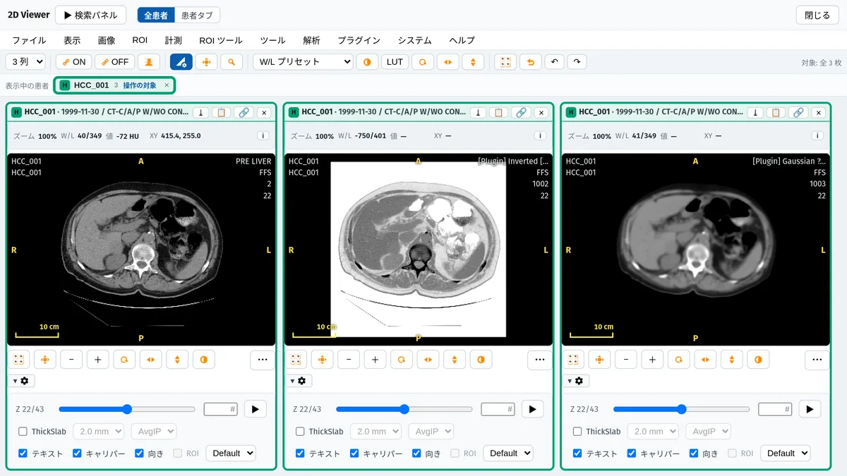
Pixel inversion, clipping and smoothing (in mm), saved as a derived series.
Apply for site support →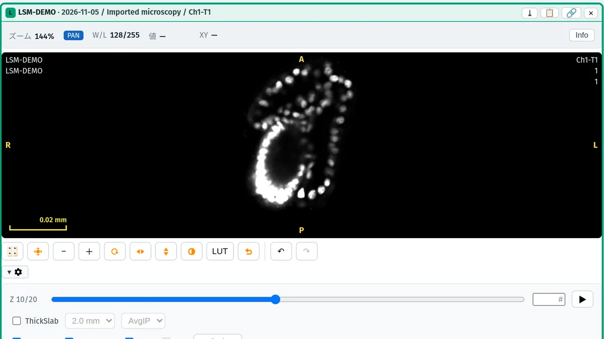
Imports Zeiss LSM and Bio-Rad PIC confocal stacks as series (desktop edition).
Apply for site support →All are for research and education only, not for diagnostic use. Plugins marked “In development” may change in timing and scope.
An Electron desktop app. Stores studies in a local H2 database and on the file system; everything from DICOM reception to the full viewer works offline.
A backend-for-frontend that connects the browser to an external PACS over DICOMweb (QIDO/WADO). Images stay on the PACS and are read from there.
GRAPHY-Next itself is free under AGPL-3.0. Onboarding, hands-on training and priority support for labs and departments are offered through the plan for companies and educational institutions.
Hours: During business hours, on an as-needed basis / Channels: Email, chat, Slack and similar (we do not offer telephone support). For details, see the support page.
On-site onboarding, hands-on training and priority support (annual agreement)
See the plan Start free instead